


Information or pictures from this site may not be used or reproduced without consent and proper citation. Please send comments or suggestions to the webmaster.
Website designed by Freckles Studio.

REAL-TIME 3D SINGLE-PARTICLE TRACKING AND 3D MULTI-RESOLUTION IMAGING
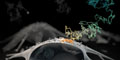
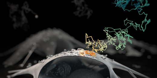
The interior of a cell is highly heterogeneous where molecular and nanoscale dynamics span multiple time and length scales. Furthermore, these dynamics all occur in three dimensions (3D). For example: How does a viral particle or a vaccine-delivery vehicle approach a living cell and become internalized? What are the dynamic molecule-level interactions that lead to a (un)successful entry? How does the intracellular cargo get delivered to its destination? To understand the molecule-nanoscale dynamics in the live-cell context, time must be explicitly considered. My lab recognized this problem very early on and has developed a real-time 3D single-particle tracking (RT-3DSPT) technology which enables us to follow a tagged nanoscale probe with 10 μs time resolution and ~10 nm spatial localization precision. We are also developing new ways to integrate this RT-3DSPT technology to a variety of imaging microscopy modalities as well as single-particle spectroscopy of a freely moving nanoparticle such as the measurement of single-particle 3D reorientation dynamics.
3D Real-Time Multi-Resolution Microscopy
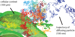
We pioneered 3D real-time multi-resolution microscopy. It is achieved by integrating, on the hardware level, the high-resolution RT-3DSPT (see below) with a lower-resolution imaging modality, taken concurrently in real time, that gives the surrounding larger environmental contexts. With this we were able to directly monitor chemical dynamics at interfaces, for example.
Links: Opt. Lett., 32, 2729-2731 (2007): conceptualization and initial realization; Nat. Nanotechnol., 9, 198-203 (2014): concurrent 3D contextual imaging realization; Faraday Discuss., 184, 359-379 (2015): technical review; J. Microbiol. Immunol. Infect., 56, 257-266 (2023): tracking individual viruses.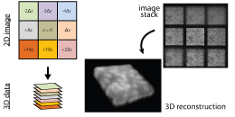
3D Multi-focal Volumetric Imaging at Video Rates
We have implemented a spatial light modulator (SLM) based multi-color, multi-focal microscope (MFM) to acquire 3D volume images at video rates (> 20 frames/second). We are currently using this to understand diffusion in confined environments.
Links: PLoS ONE, 15, e0230217 (2020): new approach to achieve uniform intensity; J. Opt. Soc. Am. B, 38, 2792-2798 (2021): MFM localization precision; Proc. SPIE, XXVIII, 11649OP (2021): SLM-MFM optimization algorithm; Sci. Rep., 12, 26343 (2022): multi-color MFM.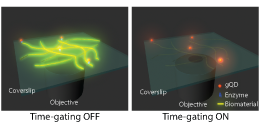
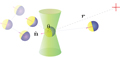
Real-Time 3D Single-Particle Tracking & Spectroscopy
We pioneered real-time 3D single-particle tracking and have since been continually furthering the concept both technically and theoretically. Most recently, we have implemented hardware-based lifetime gating to achive much improved signal-to-background ratio.
Links: J. Chem. Phys., 155, 164201 (2021): lifetime-gated RT-3DSPT.Updated: 2024.02.23.